Microscopic self-assembly of liquid crystal in Pfizer Comirnaty Covid-19 injection and dental anaesthetic.
Hi guys,
Lots of interesting reading and microscopy this week with the addition of two modifications to Karl’s scope - a powerful ring LED above light source and yesterday the use of 100W LED light source from below. Both these have provided new insights into what we are looking at in the blood and medications.
(We are writing a comprehensive document that will discuss these findings in detail along with the supporting literature… )
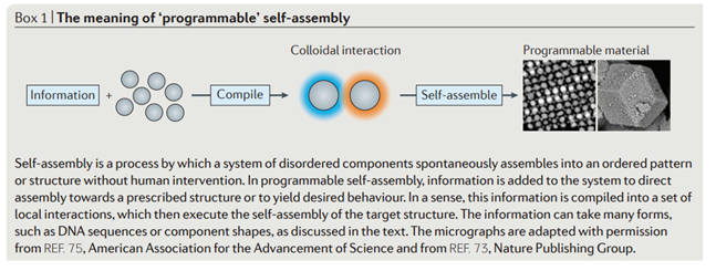
But in a nutshell this table from a 2016 review article:
The technology that supports the idea of DNA coated colloidal molecules being programmed to make complex microscopic structures also indicates that a drop in temperature can cause liquid crystal formation.
Furthermore according to 2012 article by Yonggang and colleagues
Self-assembly of nucleic acids (DNA and RNA) provides a powerful approach for constructing sophisticated synthetic molecular structures and devices (1–31). Structures have been designed by encoding sequence complementarity in DNA strands in such a manner that by pairing up complementary segments, the strands self-organize into a prescribed target structure under appropriate physical conditions (1).
From this basic principle, researchers have created diverse synthetic nucleic acid structures (27–30) such as lattices (4, 6, 8–10, 25), ribbons (15), tubes (6, 15, 25, 26), finite two dimensional (2D) and 3D objects with defined shapes, and macroscopic crystals.
Some of the most memorable videos that I have taken are from October 2022 and are on my website - please click the image below:
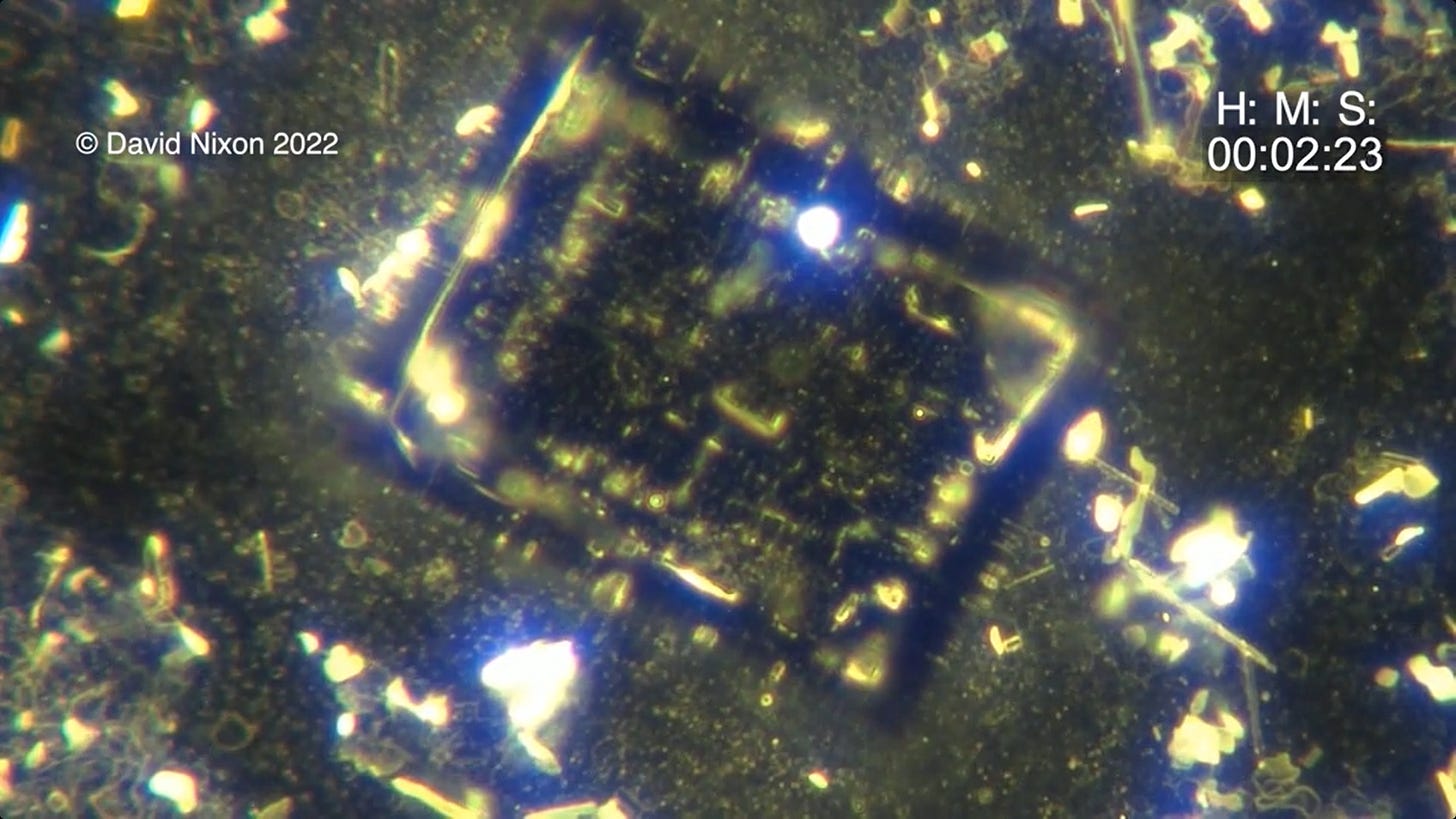
I have this video available at various speeds and I hope to make it available for download soon from the website. The particles that are shown are probably colloidal molecules coated in DNA (?). Clearly the central crystal appears to be in a liquid and the way it disappears at the end is quite something. I do not have an explanation for what appears to be structural tech surrounding the crystal.
This is a 2 minute edit of a liquid crystal forming in a less commonly used dental anaesthetic without a coverslip at approximately 200x magnification with a modest green filter.
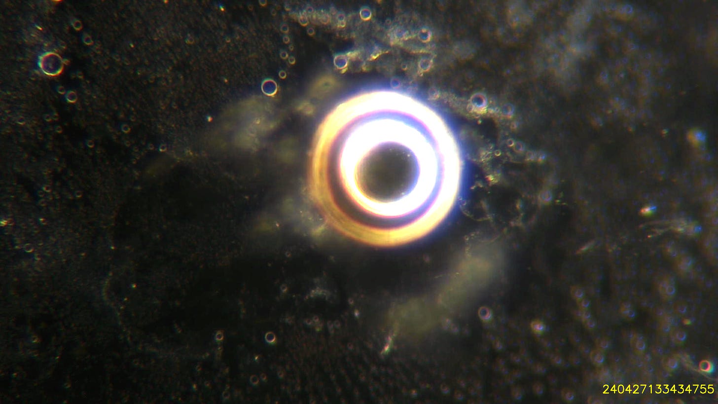
The crystal can just be seen starting at approx 10 o’clock.
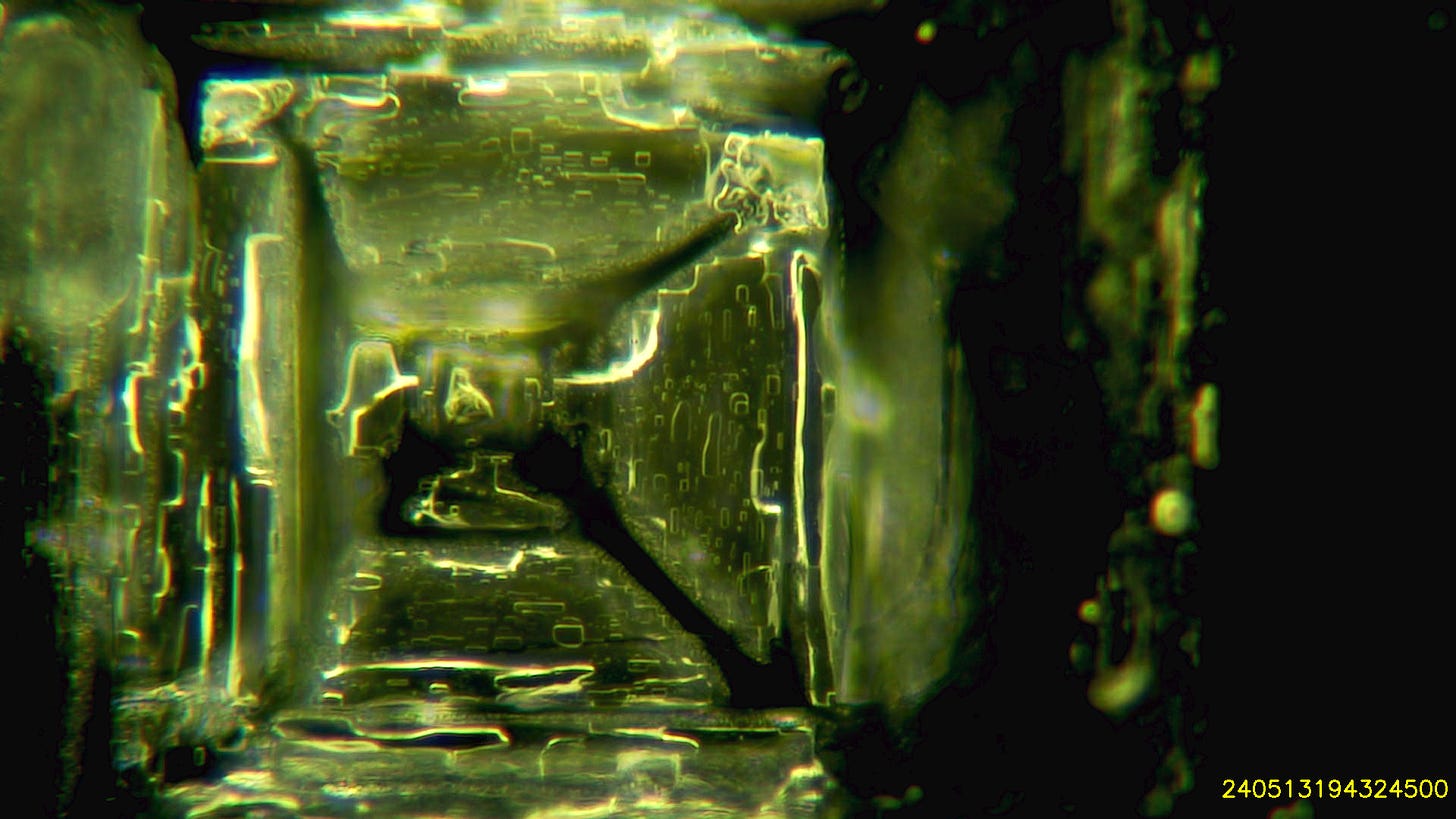
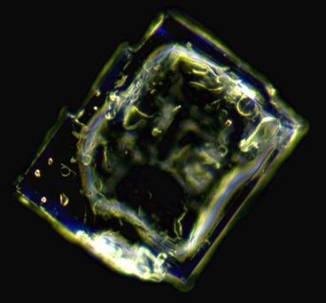
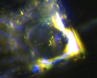
This is a close up of the crystal:
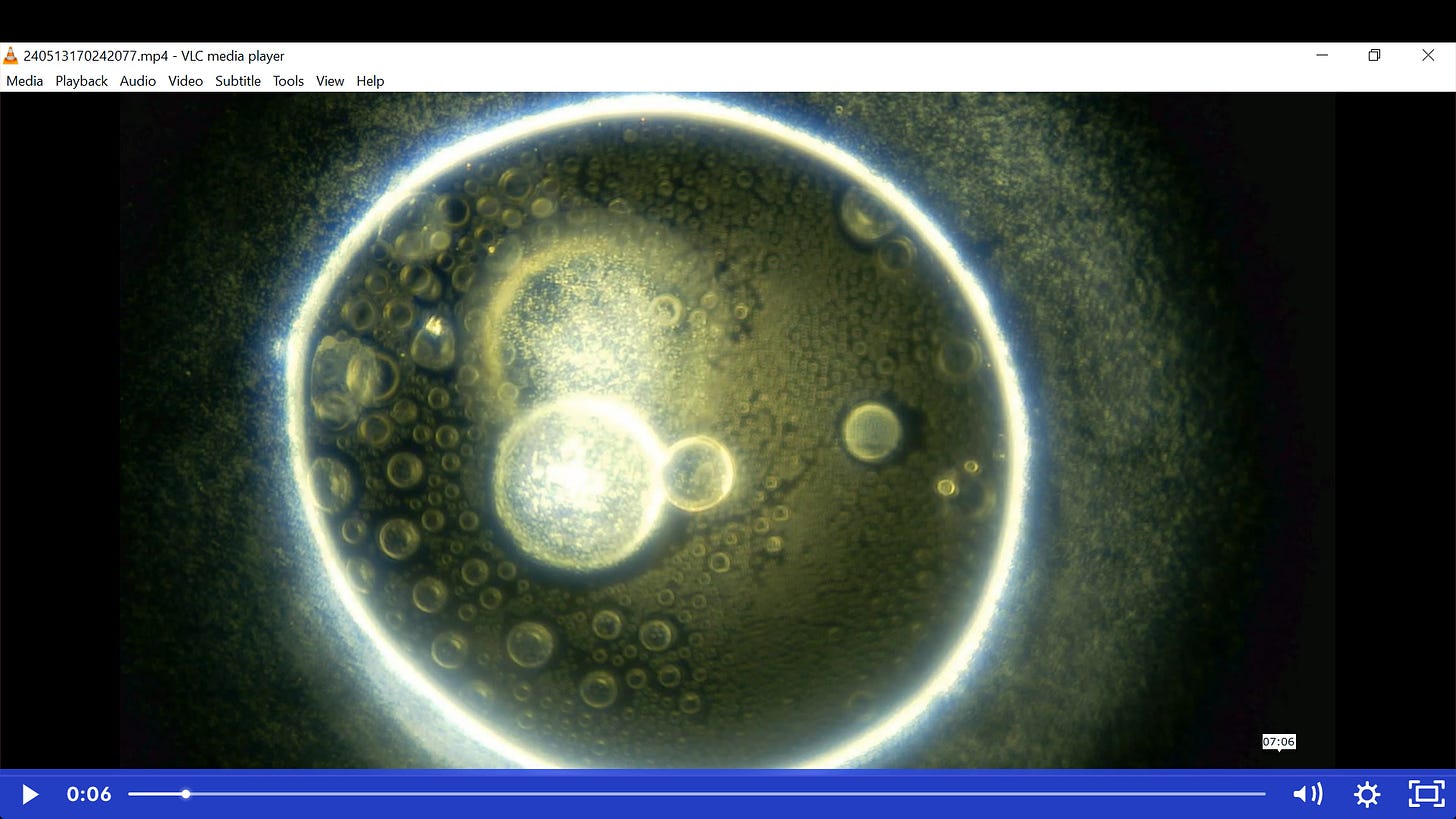
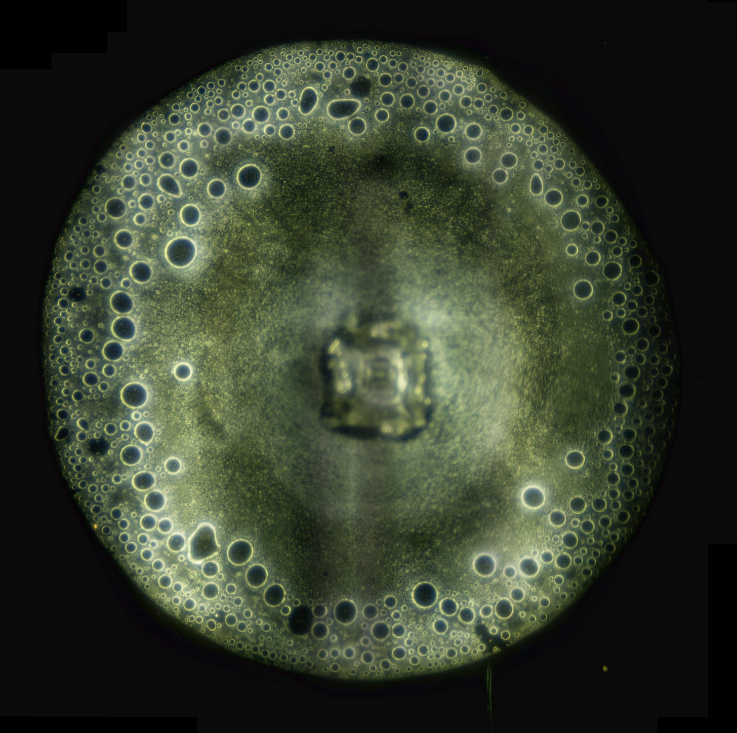
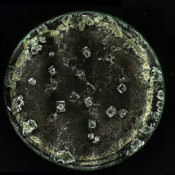
This is a photo-stitch showing the whole drop but focusing on the cells and colloidal material that have formed from the earlier processes.
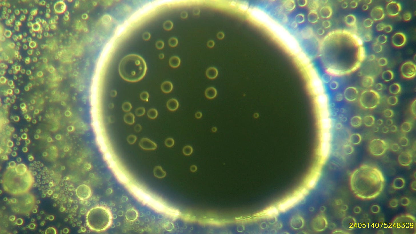
Here is a close-up of one of the cells:
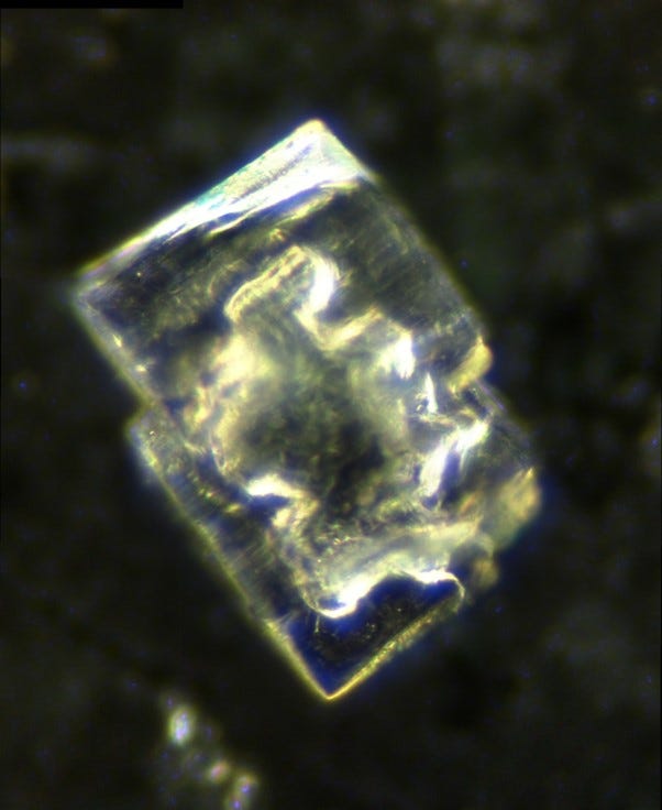
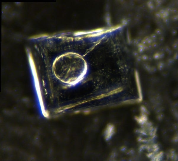
Here is a crystal from a different local anaesthetic showing a ?hydrogel bubble on top of the crystal:
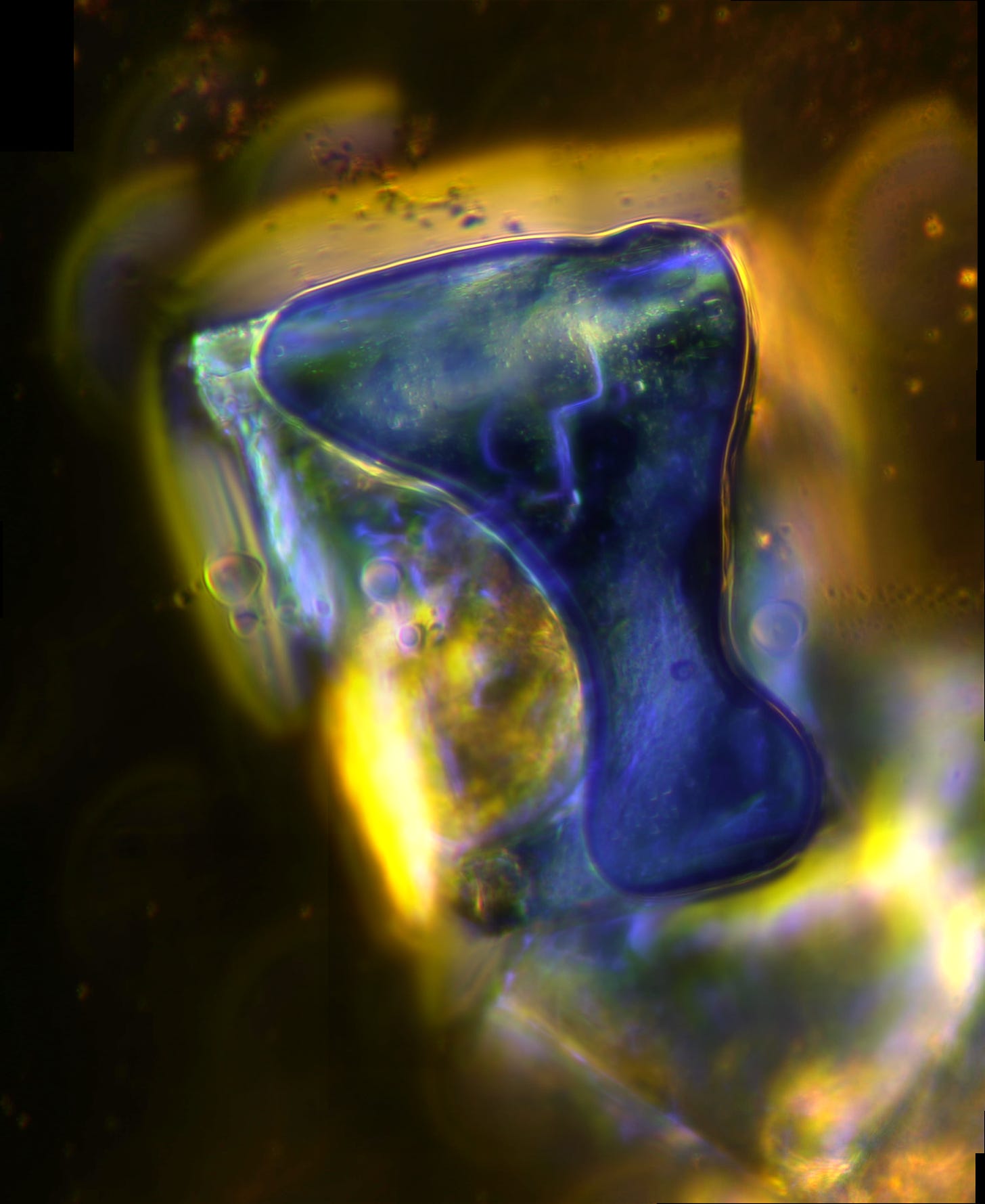
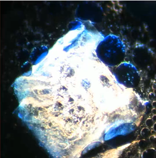
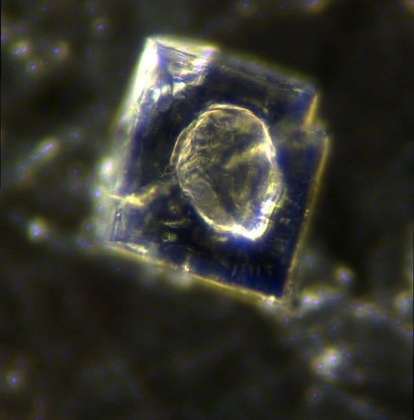
and here is a crystal from a different brand of lignocaine with a ?hydrogel bubble on top that has stained blue with crystal violet dye. The crystal has stained with fluorescein. (I have posted this one before but it is worth showing again!)
Here is a closer image from a different crystal in the same sample:
And here is a short video with crystal violet dye and lignocaine showing the cells stained blue:
Here is a photo-stitch of a drop of one of this years’ flu-shots showing a similar phenomenon with ?hydrogel on top of the crystals:
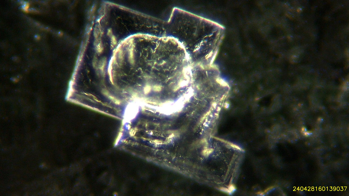
and here is a close up at 200x magnification of some of these crystals:
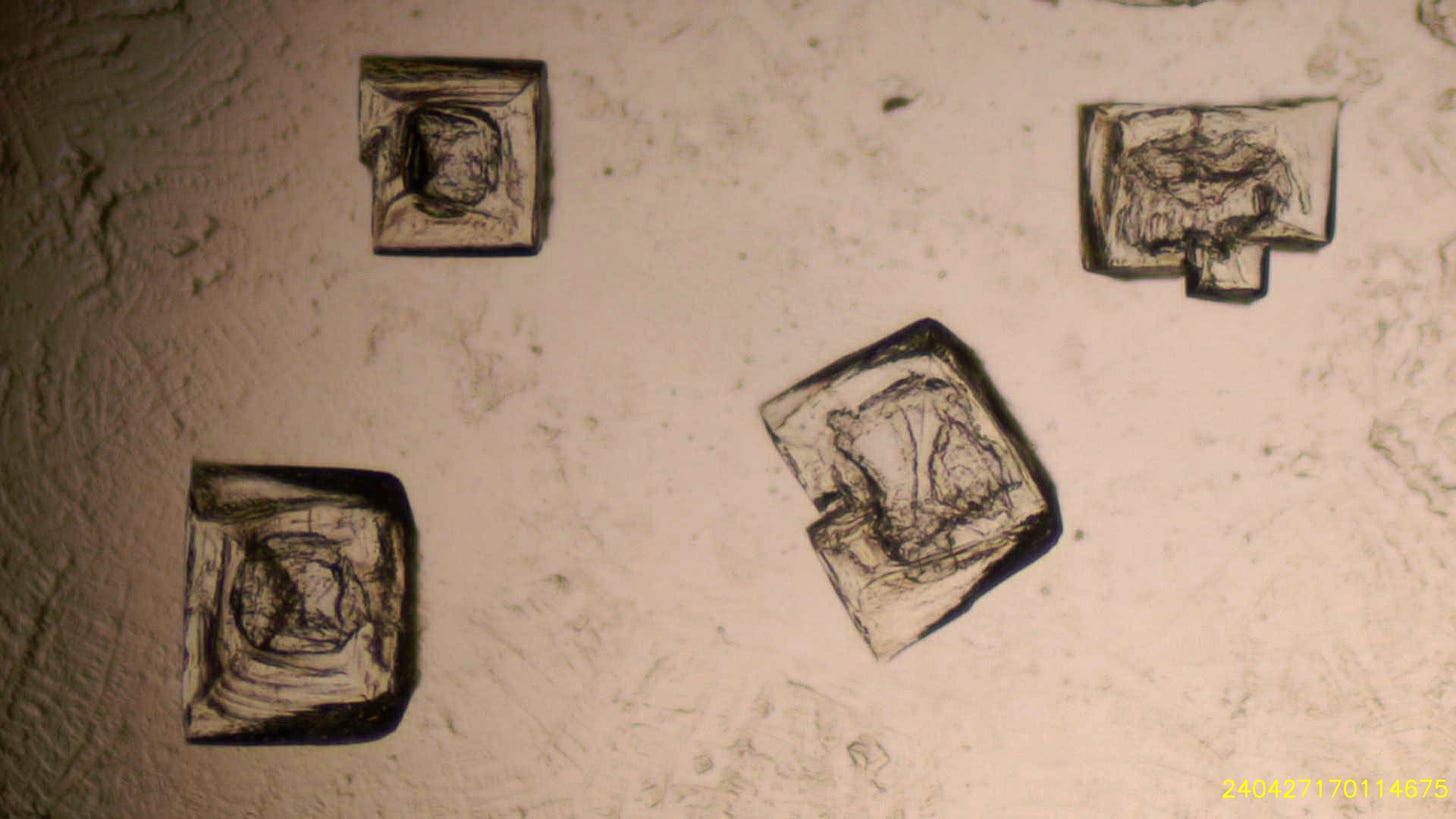
Here are four crystals in bright field, at lower magnification.
When a further drop of sample is added the crystal dissolves quickly, clouds of particles are released and the bubble on top clearly changes into a different cell structure with very thick membranes.
By matching the observations with the the relevant literature we are beginning to have a basic understanding of the processes involved. We don’t believe that there is ANY satisfactory explanation as to why this complex chemistry is present in these medications.
Our research is full-time and ongoing. We continue due to the generosity of subscribers and donors and we thank you for your support. Any contribution is gratefully received. Please consider a paid subscription and joining our weekly conversations.
Best wishes
David
All further assistance much appreciated!



No comments:
Post a Comment